
GPs can start treatment without radiographic confirmation in patients with early hip osteoarthritis.
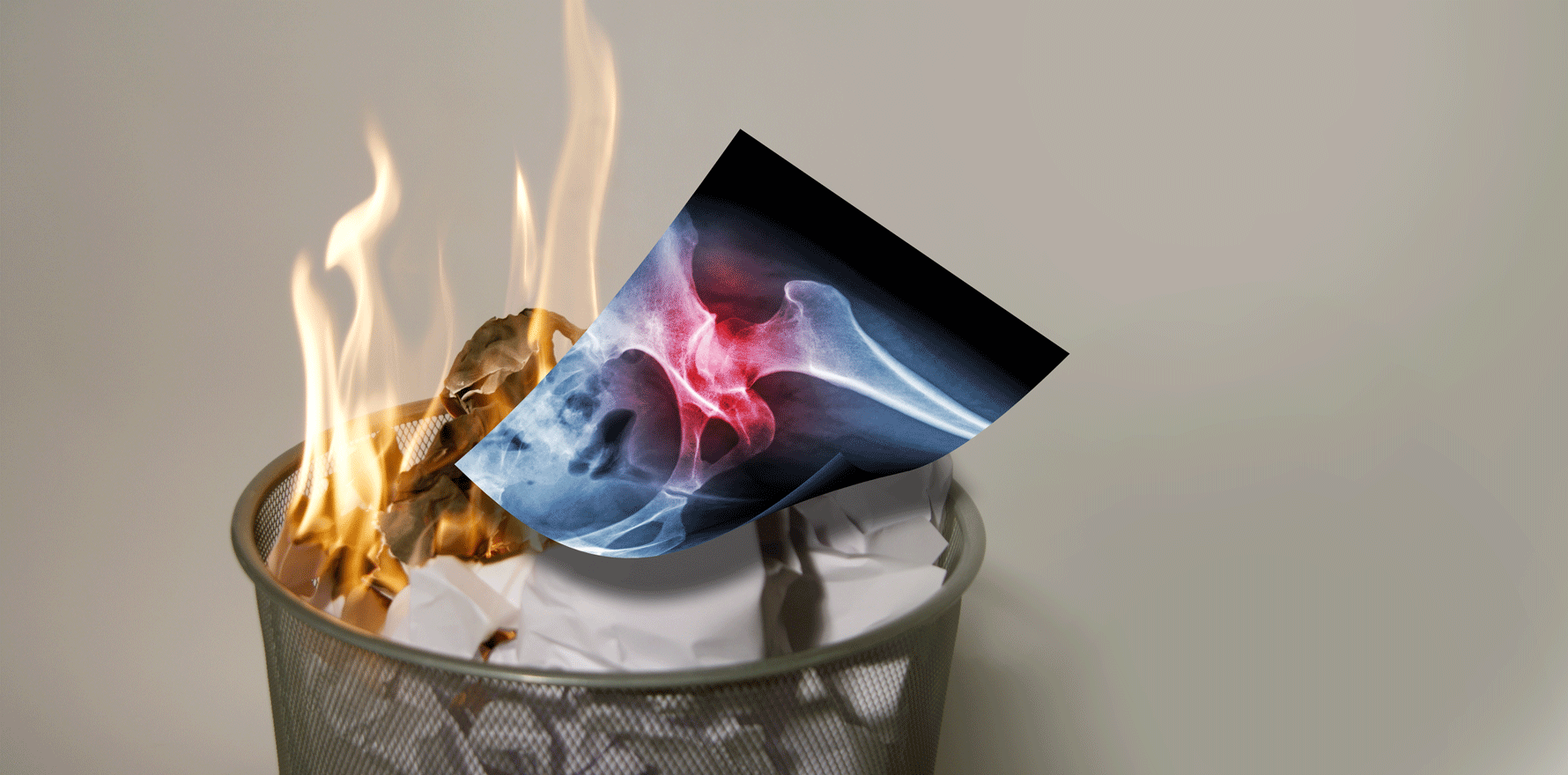
Radiographic validation of clinically suspected hip osteoarthritis has limited benefits and is not necessary for an accurate diagnosis, according to a recent study in the BJGP.
Researchers observed a modest association between hip pain and hip radiographic osteoarthritis in patients with early symptomatic hip osteoarthritis, suggesting that radiographs provide minimal assistance in identifying the condition.
To examine the association between hip pain and hip radiographic osteoarthritis, Dutch researchers analysed results from a cohort, aged between 45 and 65 years, who had hip and/or knee pain.
They found that the overall prevalence of definite hip radiographic osteoarthritis was 11%, with prevalence in painful and pain-free hips 13.3% and 9.5%, respectively. Overall prevalence of early-stage hip osteoarthritis was 35.3%, while the prevalence of painful hips was 41.2% and pain-free hips was 31.4%.
“As most people with painful hips did not have hip radiographic osteoarthritis and the association between hip pain and radiographic osteoarthritis was only moderate, radiographs likely do not help in the identification of patients with hip osteoarthritis in primary care,” the authors wrote.
“This suggests that prior guideline recommendations, mostly based on knee osteoarthritis data, which state that radiographic evidence was not required for an osteoarthritis diagnosis, apply for hip osteoarthritis as well.”
Clinical criteria of hip osteoarthritis is sufficient to commence guideline-recommended conservative osteoarthritis treatment, they said.
“A GP could – without radiographic confirmation of osteoarthritis – consider these patients as having early osteoarthritis and initiate appropriate treatments, such as education, exercise and weight loss,” they wrote.
“Therefore, referral for radiography will not likely change clinical decision making in these patients.”
Professor David Hunter, rheumatology clinical researcher at the University of Sydney, told The Medical Republic that history and clinical examination should be relied on, as opposed to routine imaging, in the majority of patients.
“If there is a high index of suspicion that an important differential exists (e.g. avascular necrosis, pigmented villonodular synovitis, osteochondritis dissecans), then that is the role that imaging might play at a diagnostic stage,” said Professor Hunter.
“It is important to recognise that these are unusual diagnoses and that most other common hip problems (e.g. gluteal tendinopathy, iliopsoas tendinitis, referred pain) should be readily identifiable based on clinical findings.”
Professor Flavia Cicuttini, head of rheumatology at Alfred Hospital, said that x-rays could be useful if, after the clinical presentation and examination, “there is the diagnosis of hip osteoarthritis and there is ongoing severe pain despite treatment”.
“In general, imaging is not needed unless there is a concern about other causes of sudden, severe onset of pain such as a fracture or other less common problems.”
While a heavy emphasis was still placed on imaging to diagnose osteoarthritis, Professor Hunter pointed out that over-imaging had harmful effects for both the patient and healthcare system, such as unnecessary costs and invasive interventions.
Overreliance on imaging may also reduce patients’ adherence to exercise and other interventions, he added.
Professor Hunter encouraged physicians to “listen to patients and engage in a discourse that facilitates participation and does not overly discourage them”.
