


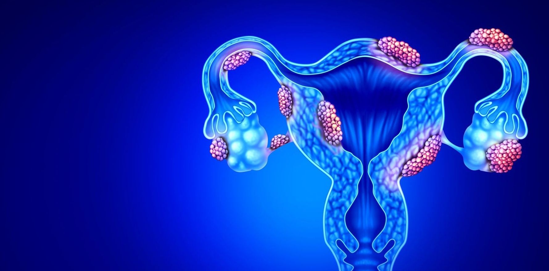
A new AI tool can reliably identify two key indicators of advanced endometriosis in pelvic scans in 18 milliseconds. But it still has a long way to go.
Researchers say EndoFusion, a new Australian AI tool, is an “initial proof of concept” that can help clinicians more accurately diagnose endometriosis without the need for laparoscopy.
The study, published in Artificial Intelligence in Medicine, found EndoFusion was more reliable than all competing models in distinguishing endometriosis-positive scans from negative ones, researchers said.
“We looked at the how well EndoFusion was able to distinguish between positive and negative cases of endometriosis and found it was able to provide a correct diagnosis 83% of the time,” said lead author Dr Yuan Zhang, from the Adelaide University’s Robinson Research Institute and the Australian Institute for Machine Learning.
According to the paper’s own results, a simpler baseline model that used no knowledge distillation outperformed EndoFusion on both ultrasound-based tasks – though the researchers’ model substantially outperformed that same baseline on the MRI tasks.
However, the AI framework remains in the early stages of development, with testing limited to small, single-site cohorts.
Non-invasive imaging modalities, including transvaginal ultrasound (TVUS) and magnetic resonance imaging (MRI), have distinct strengths in detecting signs of endometriosis, such as obliteration of the pouch of Douglas (POD) and bowel nodules (BN).
POD is often more accurately detected on TVUS than on MRI, while BN are more visible on MRI than on TVUS, the researchers reported.
However, patients typically receive only one imaging modality – often TVUS as first-line imaging.
With average diagnosis times spanning up to seven years and requiring costly surgical procedures, “no patient should be disadvantaged by being scanned with a less optimal modality for a particular endometriosis sign,” the report read.
Study co-lead Associate Professor Jodie Avery, research co-lead for Chronic Reproductive Conditions at Adelaide University’s Robinson Research Institute, said the project aimed to “democratise” endometriosis by improving access for women in rural and remote communities and those unable to afford private health care.
“Some patients could be disadvantaged if they are scanned by the less optimal option for their particular signs. Some of the imaging tools also rely on operator experience and can be costly,” Professor Avery said.
“We also want to be able to make it so that GPs are the ones who initiate this diagnostic process, even before they consider sending a patient to a gynaecologist, which in the public system is a two-year wait. Then there’s another two-year wait to have surgery,” she told The Medical Republic.
Researchers described EndoFusion as an unpaired, multimodal, multilabel learning framework, developed from IMAGENDO, an AI-driven research project led by the University of Adelaide, that enabled detection of POD and BN using both TVUS and MRI imaging data.
Related

“We’ll be able to overlay the algorithm on the information we gain on a TVUS to value-add any kind of information we might gain from an MRI as well. We’ll be able to do the reverse as well with MRIs, especially in the case of a young woman,” Professor Avery told TMR.
The researchers trained and tested their model using four separate datasets. Nearly 9000 pelvic MRI scans from the South Australian Medical Imaging service, collected over 10 years, were used to pre-train the model’s image-reading capability.
The diagnostic task, however, relied on far smaller cohorts – 171 MRI scans from 171 women, of whom 63 had POD obliteration, and only 11 had BN.
Two distinct TVUS datasets, collected from a private ultrasound clinic in Sydney, contributed 749 unique patients, including 103 POD obliteration cases and 116 patients with 13 BN cases.
Critically, no patient contributed both an MRI and a TVUS scan, a gap the researchers addressed by incorporating a label-based pairing technique – pairing scans from different patients that displayed the same findings.
They acknowledged, however, that because the paired scans never came from the same patient, some of EndoFusion’s apparent gains may instead reflect how the scans were matched, rather than the model truly learning to read across both imaging types.
Dr Yuan Zhang told TMR the paper had been written two years prior and that researchers now had access to a much larger dataset.
“Once we organise all the new data coming in, we will retrain the model, and we can expect a much better performance,” she said.
The researchers also employed Dynamic Mutual Knowledge Distillation (DMKD), using a worst-student-oriented teacher-selection strategy during AI model training.
After each training round, whichever imaging modality performed worst on validation checks was designated the “student” for the next round, while the better-performing modality became the “teacher”.
DMKD achieved the best overall discrimination performance, particularly for MRI-based POD and BN classification, with area under the curve (AUCs) of 0.765 and 0.903, respectively.
While AUC does not directly translate to real-world diagnostic accuracy, the model was more reliable than any comparison method in distinguishing positive from negative across MRI and TVUS combined.
Associate Professor Jodie Avery said researchers were still working out the best way to implement it as a clinical tool.
One proposal was a plug-in that radiology practices could use to report on MRIs and TVUS scans, integrated into their existing software. Another was for it to form part of a gynaecological practice package, she told TMR.
“We’re sort of tossing up a couple of different ideas on how to roll this out so it can be translated and get out to people in primary healthcare,” she said.
The research could offer insights into other diseases facing similar multimodal, multilabel imaging classification challenges, including other gynaecological disorders, prostate cancer, breast cancer and foetal abnormalities, researchers reported.
Professor Avery told TMR further TVUS data from patients in the existing study was being collected to identify additional endometriosis markers, including endometriomas and uterosacral ligament involvement.
Professor Avery said a prospective cohort study of women aged 14 to 25 years was underway, with GPs in South Australia able to refer young women with pelvic pain to take part and receive follow-up for the next three years.
The research was conducted in collaboration with Flinders University, Benson Radiology, Omni Ultrasound and Gynaecological Care, the University of Surrey, McMaster University Medical Centre, and Mohamed bin Zayed University of Artificial Intelligence.
Read the full report here.
