Diagnostic uncertainties require a logical and pragmatic approach – and an open mind.
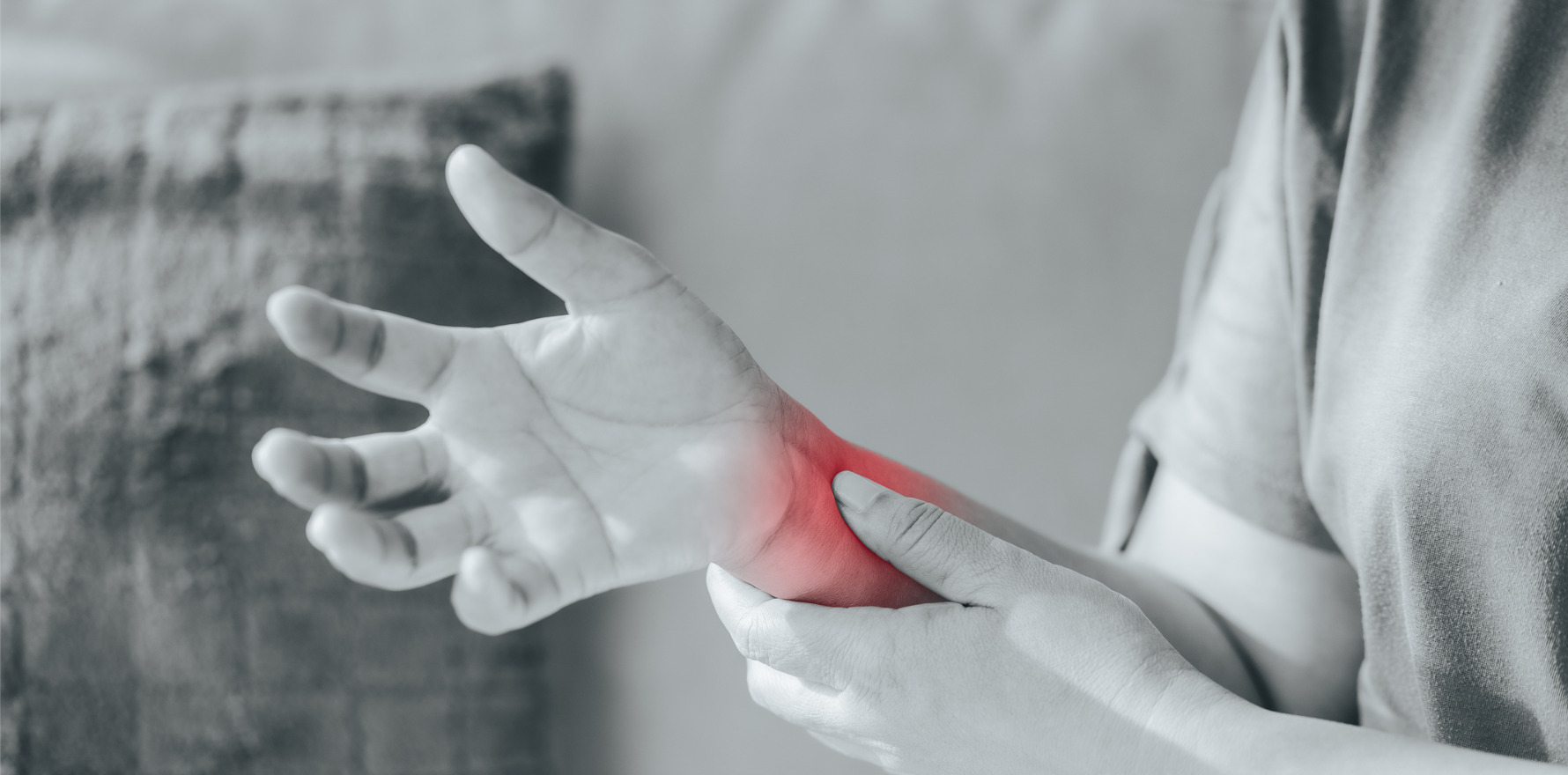
An 80-year-old female presents with acute right-wrist swelling, increasing in severity over the course of 24 hours.
She was reviewed one week after the onset of pain. The pain has been waking her at night, there is restricted wrist movement and difficulty performing daily activities. Other joints are unaffected. There was no preceding trauma or infection, and she has been afebrile.
She has a history of ovarian cancer requiring hysterectomy and salpingo-oophorectomy five years prior with adjuvant chemotherapy. There has been no evidence of relapse.
She has stage III chronic kidney disease with estimated GFR of 37. This is secondary to hypertension, for which she takes candesartan 16mg daily, hydrochlorothiazide 25mg daily, atenolol 50mg daily and lercanidipine 20mg daily. She has type 2 diabetes requiring metformin 1g daily with excellent control of HbA1c. There is a history of gout, for which she takes allopurinol 300mg daily. There have been no recent attacks of gout. She has rate-controlled atrial fibrillation and is taking apixaban 2.5mg daily.
Examination shows a grossly swollen right wrist with synovitis. It is very tender and painful to move. There is puffiness of the fingers and dorsum of the hand, but there did not appear to be definite synovitis. Examination of other joints shows evidence of nodal osteoarthritis in her fingers and osteoarthritis of the base of the thumb. There is no synovitis elsewhere and no psoriasis or nail changes.
Initial blood tests show creatinine 117 with eGFR 37, serum uric acid 0.38mmol/L, CRP 26 (N<5) and ESR 63 (N<21). There is mild normochromic normocytic anaemia with haemoglobin 105, presumed to be secondary to renal impairment.
The potential differentials are broad.
The most common cause of acute wrist swelling in the elderly is acute pseudogout. This is due to calcium pyrophosphate (CPPD) crystals causing an acute inflammatory response. She does have a history of gout but is adherent with allopurinol and has not had recent attacks. Gout attacks can occur in the wrist, but this is unusual except in poorly controlled gout. In addition, all her previous episodes of gout have been in her feet, and her serum uric acid has been relatively well controlled, although it is marginally above the target serum uric acid of 0.36mmol/L.
She is on low-dose anticoagulation and a haemarthrosis is possible. Renal impairment also impairs platelet function and increases bleeding risk. Infection should also be considered in a monoarthritis, especially given her risk factors of age, diabetes and renal impairment.
Psoriatic arthritis would be less likely given there is no history of psoriasis, but psoriatic arthritis still occurs in 10% of people without psoriasis. This could also be an initial presentation of a more systemic inflammatory condition such as rheumatoid arthritis.
The best test for a monoarthritis is a joint aspirate. Ultrasound showed evidence of synovitis and a small joint effusion. Only 0.5mL of blood-stained synovial fluid was obtained with an ultrasound-guided joint aspirate. It is not uncommon to aspirate only a small amount of synovial fluid even with extensive wrist swelling. Cell count was not performed on the aspirate but polarising light microscopy did not detect crystals and culture was negative.
X-rays of the hands, wrists, feet and knees were performed. The aim was to look for chondrocalcinosis as this is frequently the only way to diagnose pseudogout. The X-rays of the hands showed radiocarpal and first-CMC-joint osteoarthritis but no chondrocalcinosis. X-rays of the knees shows moderate medial-compartment osteoarthritis with chondrocalcinosis. X-rays of her feet showed a small erosion in the first MTP joint, consistent with past evidence of gout. RF and CCP antibodies are negative.
My provisional diagnosis is acute pseudogout, given the presence of chondrocalcinosis in the knee X-ray.
Chondrocalcinosis on X-ray of any joint raises the possibility of any monoarthritis being pseudogout. CPPD crystals are difficult to detect by polarising light microscopy with a sensitivity of around 50%, although this may be higher with experienced technicians. This contrasts with urate crystals, which are much easier to detect. The sensitivity of light microscopy for urate crystals is over 90%. Thus, a negative aspirate for CPPD does not exclude pseudogout and she has a radiological confirmation of chondrocalcinosis. In addition, only a tiny joint aspirate was able to be examined.
A wrist-joint corticosteroid injection was performed with injection of 40mg Depo-Medrol. This did provide initial relief. However, the inflammation returned.
She was started on prednisone 15mg daily with a taper over six weeks. She was reviewed at the end of this period and reported increased pain and swelling when she stopped prednisone. ESR was still raised at 40 (N<21) and CRP was still raised at 15 (N<5).
Pseudogout tends to be acute and self-limiting, with a course that lasts a few weeks. Her clinical course is now atypical for pseudogout. It is possible to get a “pseudo-rheumatoid” pattern with CPPD where there is chronic inflammatory synovitis, but this is more likely to be polyarticular.
Differentials would include an indolent atypical infection (not detected on previous joint aspirate) or a chronic inflammatory arthritis, although she would not meet formal criteria to diagnose rheumatoid arthritis. A synovial biopsy could help to differentiate this, although this is invasive, and it was elected not to do this after discussion with the patient and her family.
Although she responded very well to prednisone, long-term steroid use is not appropriate. Low-dose colchicine 0.5mg daily (a reduced dose given her renal impairment) was not effective. I felt she required DMARD therapy to suppress the synovitis. Chronic kidney disease does make the options more challenging.
I elected for low-dose methotrexate 10mg weekly, with close monitoring of her full blood count, electrolytes and liver function tests. This was well tolerated, and the synovitis resolved over the course of two months. No other joints were involved over the course of her disease. Her wrist synovitis remained under excellent control for a further six months and methotrexate was ceased.
This case demonstrates the frequent diagnostic uncertainty in rheumatology as well as the management challenges with co-morbidities. It is appropriate to have a logical and pragmatic approach in the face of diagnostic uncertainty, as well as keeping an open mind!
Dr Andrew Jordan is a rheumatologist based in Parramatta, Sydney, with a special interest in inflammatory arthritis, gout and PRP injections.
